Definition/Description
A quadriceps muscle strain is an acute tear of the quadriceps muscle. This injury is usually caused by severe muscle strain, often at the same time as forceful contractions or repetitive functional overloading. The quadriceps, which consist of four parts, can be overloaded by repeated eccentric muscle contractions of the knee extensor mechanism. [1] Quadriceps Injury
Acute strain injuries of the quadriceps commonly occur in athletic competitions such as soccer, rugby, and football. These sports regularly require sudden forceful eccentric contractions of the quadriceps during regulation of knee flexion and hip extension. High forces in muscle-tendon units with eccentric contractions can lead to stress injury. Excessive passive stretching or activation of overstretched muscles can also cause strain. Of the quadriceps muscles, the rectus femoris is the most frequently strained. Several factors predispose this muscle and others to more frequent stress injuries. These include muscles that cross two joints, with a high percentage of type II fibers and muscles with complex muscle architecture. Muscle fatigue has also been shown to play a role in acute muscle injury. Quadriceps Injury
Image: Quadriceps femoris muscle (highlighted in green) – posterior
Anatomy And Biomechanics
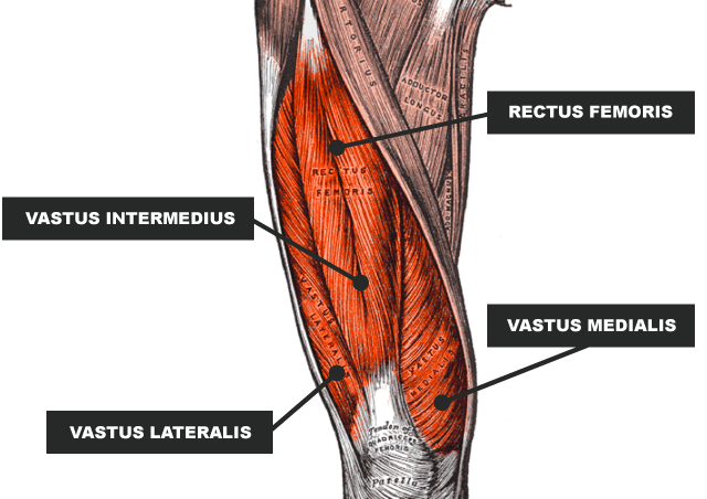
The quadriceps muscles consist of the rectus femoris, vastus lateralis, vastus medialis, and vastus intermedius (Figure 1 and Figure 1 and Figure 2 and Figure 3 and Figure 4). The rectus femoris is centrally located on the posterior thigh and has two origins (Figure 5). The more superficial fibers originate as tendons in the anterior inferior iliac spine (AIIS), while the deeper fibers arise from the acetabular rim. These two muscle segments are join by a myofascial layer, sometimes called the central tendon, that spans two-thirds of the length of the rectus femoris. The vast lateral muscle originates from the proximal femur, the lateral border, and the region of the greater trochanter (Figure 6 and Figure 7). The vast lateral (Figure 8) has the largest volume of all the quadriceps. It contributes to knee extension but also pulls the patella posteriorly.
The vast medialis is the shortest of the quadriceps and originates from the medial femur near the intertrochanteric line. As a small muscle of low mass, the vast medialis must maintain adequate strength to counteract the vast lateralis. The vast intermedius (Figure 10) originates from the proximal aspect of the femur at the superior intertrochanteric line (Figure 11). The vast intermedius helps stabilize the midline tracking of the patella during knee extension.
The joint action of the quadriceps can produce powerful knee extension. The muscle inserts on the patella as the common quadriceps tendon. This tendon then wraps around the patella and inserts into the tibial tuberosity. The part of the tendon that extends from the patella to the inferior is commonly call the patella tendon.
The quadriceps muscle is innervate by the femoral nerve (Figure 12), which arises from nerve roots in the second through fourth lumbar vertebrae (L2 through L4). The vast lateralis and vastus medialis receive a large portion of their innervation from the L3 nerve root.
HOW TO TREAT AND PREVENT A QUAD STRAIN
Historically, your healthcare provider will likely recommend rest and reduced activity. Following the RICE method (rest, ice, compression, elevation) can help reduce swelling and pain. Your doctor may also recommend over-the-counter pain relievers or anti-inflammatory medications for moderate muscle strains. For more severe quad strains, your doctor may refer you to a physical therapist to help safely regain strength and mobility during recovery.
As powerful as they are, quads are still prone to injury if neglected. Some steps you can take to prevent quad injuries include warming up properly before any activity and taking time to gently stretch and cool down your quads after exercise.
Strengthening your quads, hamstrings and hip flexors can also help reduce your risk of injury. Strong muscles are more resistant to stress and can provide more support to your joints during activity.
Tendon Weakness
A weak extensor sinew is additional seemingly to rupture. several things will cause sinew weakness.
Tendinitis. Inflammation of the extensor sinew, known as extensor tendonitis, weakens the sinew. It may cause little tears. extensor tendonitis is most typical in those who run and participate in sports that involve jumping.
Mozzie sickness. Weak tendons may be cause by diseases that disrupt the blood offer. Chronic diseases that may weaken tendons include:
- Chronic urinary organ (kidney) failure.
- Other conditions related to urinary organ chemical analysis
- Hyperparathyroidism
- Gout
- Blood cancer
- Joints
- Systemic LE (SLE)
- Diabetes mellitus
- Infection
- Metabolic sickness
Treatment for quadriceps tear or strain
The treatment protocol for a quadriceps tear or strain depends on your age, activity level, and the size of the tear.
Non-surgical treatment options for a quadriceps tear or strain:
Immobilization with a knee brace – A brace will help keep the knee straight until it fully heals, usually three to six weeks.
Physical Therapy – Physical therapy for a quadriceps tear will include specific exercises that restore range of motion and strength.
Platelet Rich Plasma (PRP) Injections – PRP therapy is a new form of therapy where the patient’s blood is taken, put through a centrifuge and then inject back into the body to facilitate healing.
People with complete quadriceps tears may need surgery to reattach the torn tendon to the knee cap. Surgery is most successful when the repair is done soon after the injury.
What are the different types of quadriceps injuries?
The most common injury to the quadriceps is a contusion or bruise, caused by a direct blow to the hamstring, which can damage some of the blood vessels within the muscle and cause bleeding. It causes pain due to inflammation of the surrounding muscles. Because the rectus femoris muscle is closest to the surface of the posterior thigh, it is the muscle most often confuse.
If there is significant bleeding into the muscle, compartment syndrome may occur, where the pressure inside the compartment containing the quadriceps rises above the blood pressure, thereby depriving the muscle tissues of oxygen-rich blood pumped by the heart. Prevents delivery.
The quadriceps can also become strained from overuse or overstretching.
Muscle strains can be classified according to the severity of the injury. There are three different grades of muscle tension. A grade 1 strain involves muscle fibers that are stretch but not torn. A grade 2 strain involves a muscle that is partially torn with more extensive damage. Grade 3 strains are when a muscle is completely torn.
Tendonitis describes inflammation of the tendon (itis = inflammation).
Jumper’s knee or peripatellar tendinosis describes a chronic degeneration of the tendon where the muscle fibers just above the patella (knee cap) transfer into the tendon fibers.
Patellar tendinitis describes inflammation under the patella on the surface of the knee joint.
Osgood-Schlatter disease, also known as apophysitis of the tibial tubercle, occurs when there is inflammation of the bone where the patellar tendon connects the quadriceps muscle to the tibia. Although it is usually see in teenagers, it can also occur in adults.
The quadriceps or patellar tendon may rupture completely, leading to an inability to fully extend or straighten the knee.
Rarely, the fascia surrounding and containing the quadriceps can tear and the defect can cause the muscle to herniate.

